SOME SPECIFICATIONS

ROI in the ascending aorta
4D-FLOW VISUALIZATION
You can open 4D-Flow DICOM files, visualize the dynamic images in a classic MPR (axial-coronal-sagittal) view with a velocity color superposition. Dynamic cursors let the user to position the ROIs following the anatomical landmarks.
A 3D angiogram visualization, including streamlines, is also available.
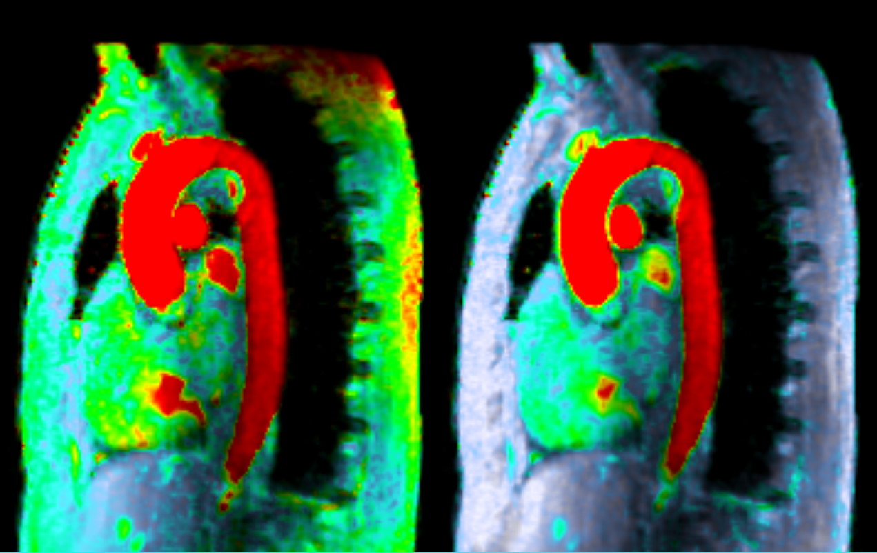
Background offset correction example
BACKGROUND OFFSET CORRECTION
Lattido includes a novel backgound offset correction algorithm that automatically corrects for Eddy currents effects on 4D-Flow images.
The algorithm is based on a recent publication of the team: Automatic Correction of Background Phase Offset in 4D-flow of Great Vessels and of the Heart in MRI Using a Third-Order Surface Model, MAGMA 2019 Dec;32(6):629-642.
doi: 10.1007/s10334-019-00765-z
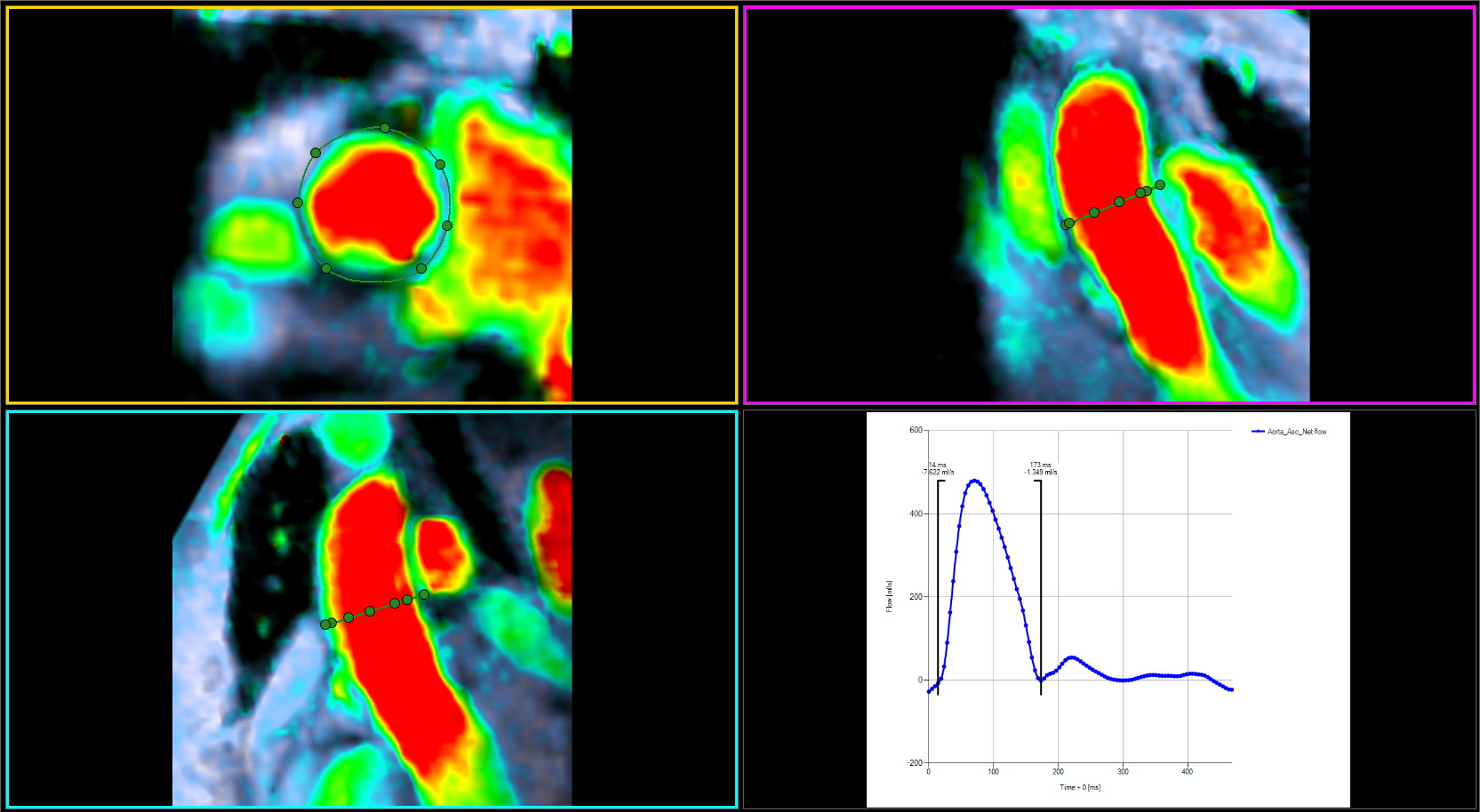
Ascending aorta blood flow estimation
BLOOD FLOW ESTIMATION
You can draw a region of interest (ROI) around a vessel or the heart in an oblique arbitrary plane and measure the positive, negative and net flow. Time cursors will let you easily select the desired range within the time phases to integrate the flow volume.
An intuitive method will let you draw a few ROIs and interpolate them through the entire cardiac cycle.
We have recently analyzed the influence of ROI size, angulation and resolution in an article published in Physiological Measurements
doi: 10.1088/1361-6579/abe525

Aortic pulse wave velocity using blood flow estimations
AORTIC PULSE WAVE VELOCITY
Using blood flow estimations along the thoracic aorta, the velocity of a pulse wave propagation can be estimated as a surrogate of
regional aortic stiffness. In the following article published in the International Journal of Cardiology, we have analyzed the
influence of ascending aorta dilatation on aortic elasticity evaluating bicuspid and tricuspid patients
doi: 10.1016/j.ijcard.2020.11.046
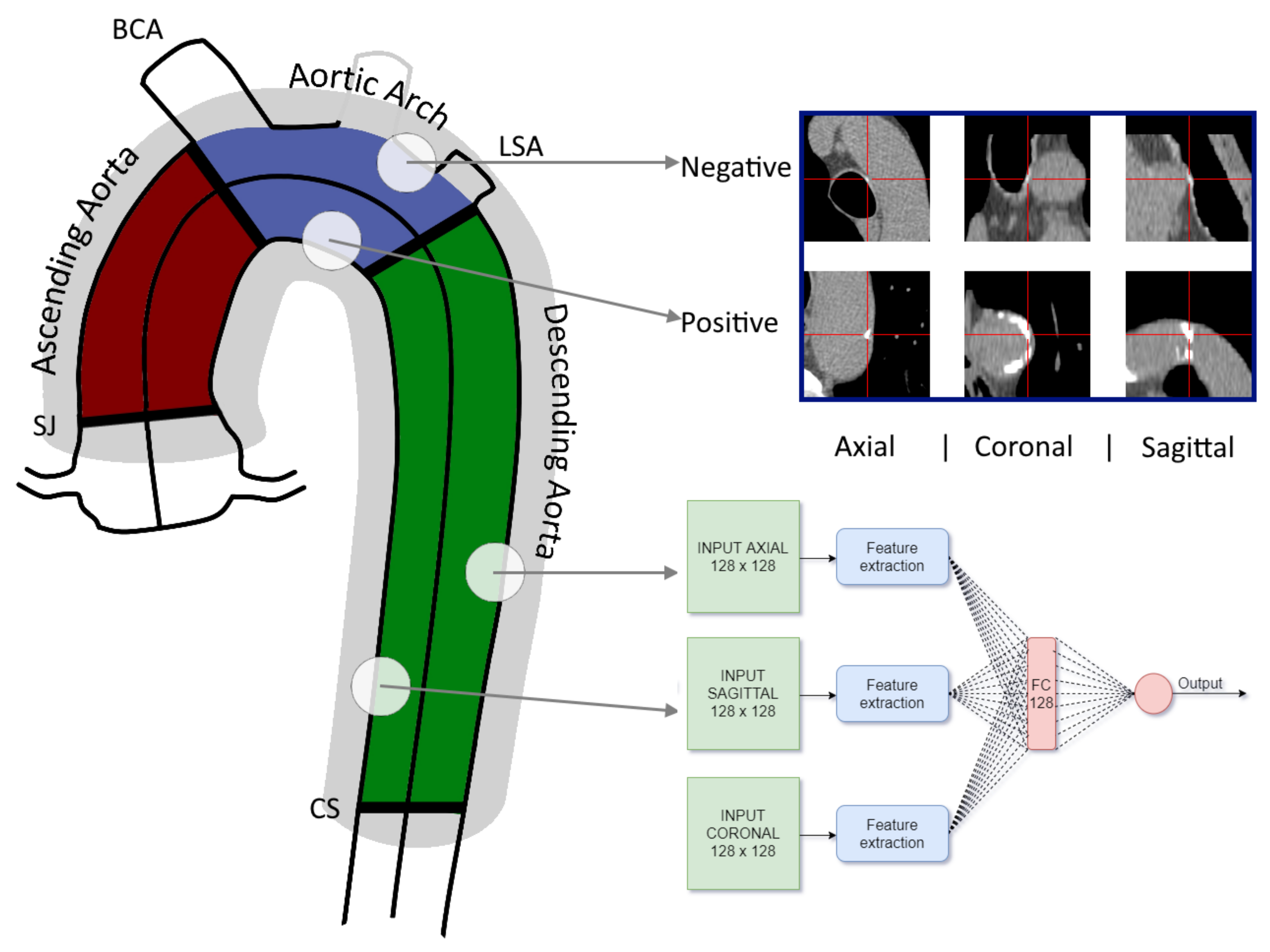
Aortic calcium detection using convolutional neural networks and Lattido
THORACIC AORTA CALCIUM DETECTION USING CNNs
Arterial calcification is an independent predictor of cardiovascular disease events whereas thoracic aorta calcium detection might anticipate extra-coronary outcomes. In this work, we trained six convolutional neural networks (CNNs) to detect aortic calcifications and to automate the TAC score assessment using Lattido. A CNN that combined axial and sagittal patches depending on the candidate aortic location ensured an accurate TAC score prediction.
doi:10.3390/tomography7040054